MOSAIC 2016 – ImageJ plugin for Biological Fluorescence Microscopy

MOSAIC 2016

:: DESCRIPTION

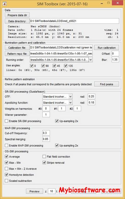
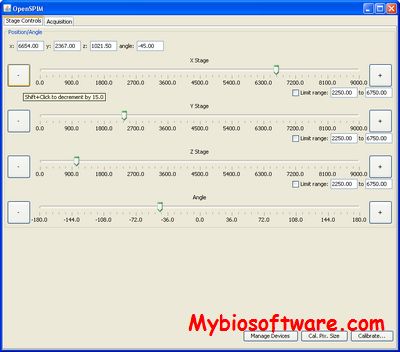
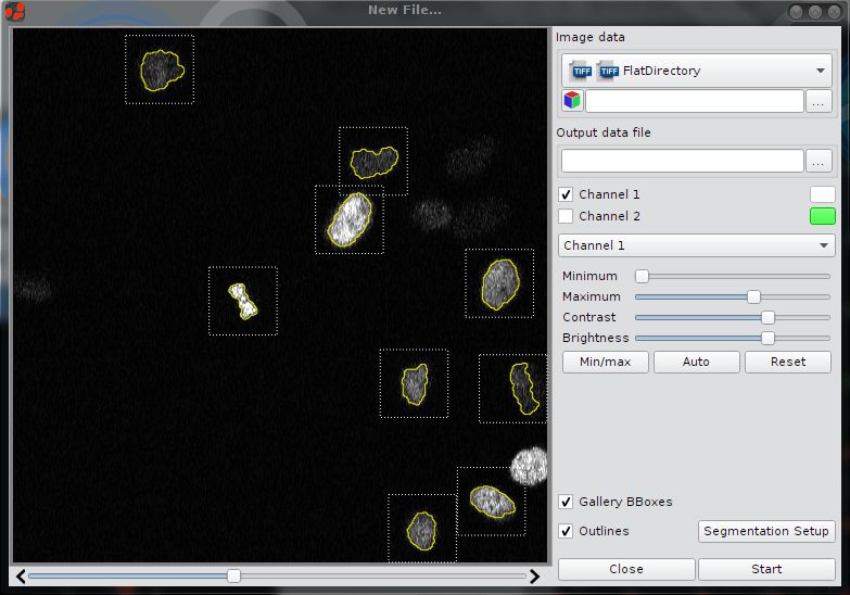
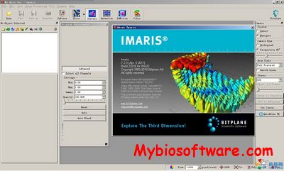
MOSAIC (MOdels, Simulations, and Algorithms for Interdisciplinary Computing) , the image-processing algorithms developed at the MOSAIC Group for fluorescence microscopy , are available as plugins for the popular free image processing software ImageJ.

::DEVELOPER

MOSAIC group

:: SCREENSHOTS

N/A

:: REQUIREMENTS

:: DOWNLOAD

 MOSAIC

:: MORE INFORMATION

Citation:

I. F. Sbalzarini and P. Koumoutsakos.
Feature Point Tracking and Trajectory Analysis for Video Imaging in Cell Biology,
Journal of Structural Biology 151(2):182-195, 2005.

Xmipp 3.1 – X-Windows-based Microscopy Image Processing Package

Xmipp 3.1

:: DESCRIPTION

Xmipp is a suite of image processing programs, primarily aimed at single-particle 3D electron microscopy.

::DEVELOPER

Biocomputing Unit

:: SCREENSHOTS

:: REQUIREMENTS

  •  Windows with Cygwin /Linux/MacOsX
  • Python

:: DOWNLOAD

 Xmipp

:: MORE INFORMATION

Citation

de la Rosa-Trevín JM, Otón J, Marabini R, Zaldívar A, Vargas J, Carazo JM, Sorzano CO.
Xmipp 3.0: an improved software suite for image processing in electron microscopy.
J Struct Biol. 2013 Nov;184(2):321-8. doi: 10.1016/j.jsb.2013.09.015. Epub 2013 Sep 26. PMID: 24075951.

C.O.S. Sorzano, R. Marabini, J.M. Carazo, J. Velazquez-Muriel, J.R. Bilbao-Castro, S.H.W. Scheres, J.M. Carazo and A. Pascual-Montano.
XMIPP: A new generation of an open-source image processing package for Electron Microscopy.
J Struct Biol. 2004 Nov;148(2):194-204.

SIMToolbox 2.12- MATLAB toolbox for Structured Illumination Fluorescence Microscopy

SIMToolbox 2.12

:: DESCRIPTION

SIMToolbox is an open-source, modular set of functions for MATLAB designed for processing data acquired by structured illumination microscopy.

::DEVELOPER

Multimedia Technology Group

:: SCREENSHOTS

SIMToolbox

:: REQUIREMENTS

  • Windows
  • MatLab

:: DOWNLOAD

 SIMToolbox

:: MORE INFORMATION

Citation

SIMToolbox: a MATLAB toolbox for structured illumination fluorescence microscopy.
Křížek P, Lukeš T, Ovesný M, Fliegel K, Hagen GM.
Bioinformatics. 2015 Oct 6. pii: btv576.

OpenSPIM 20150427 – Open-access Light-sheet Microscopy Platform

OpenSPIM 20150427

:: DESCRIPTION

OpenSPIM is an Open Access platform for applying and enhancing Selective Plane Illumination Microscopy (SPIM).

::DEVELOPER

OpenSPIM team

:: SCREENSHOTS

OpenSPIM

:: REQUIREMENTS

  • Windows / Linux / MacOSX
  • Java

:: DOWNLOAD

 OpenSPIM

:: MORE INFORMATION

Citation

Nat Methods. 2013 Jul;10(7):598-9. doi: 10.1038/nmeth.2507. Epub 2013 Jun 9.
OpenSPIM: an open-access light-sheet microscopy platform.
Pitrone PG, Schindelin J, Stuyvenberg L, Preibisch S, Weber M, Eliceiri KW, Huisken J, Tomancak P.

imageHTS 1.43.0 – Analysis of high-throughput Microscopy-based Screens

imageHTS 1.43.0

:: DESCRIPTION

imageHTS is an R package dedicated to the analysis of high-throughput microscopy-based screens. The package provides a modular and extensible framework to segment cells, extract quantitative cell features, predict cell types and browse screen data through web interfaces.

::DEVELOPER

Huber Group

:: SCREENSHOTS

N/A

:: REQUIREMENTS

  • Linux/ WIndows/MacOsX
  • R package
  • BioConductor

:: DOWNLOAD

  imageHTS

:: MORE INFORMATION

CellCognition Explorer 1.0.2 – Analysis of Cellular Phenotypes Images in Microscopy

CellCognition Explorer 1.0.2

:: DESCRIPTION

CellCognition Explorer is an open-source image processing tool for the analysis of cellular phenotypes in microscopy. CellCognition Explorer enables phenotype classification by supervised machine learning. To detect rare phenotypes, outlier morphologies can be automatically found by novelty detection methods. A key feature of CellCognition Explorer is an improved classifier training procedure based on automated pre-processing of the full data set into cell gallery images, which can be automatically sorted based on phenotype similarity for efficient iterative classifier training.

::DEVELOPER

CellCognition Explorer team

:: SCREENSHOTS

:: REQUIREMENTS

  • Windows / Linux / MacOsX

:: DOWNLOAD

CellCognition Explorer

:: MORE INFORMATION

Citation

Sommer C, Hoefler R, Samwer M, Gerlich DW.
A deep learning and novelty detection framework for rapid phenotyping in high-content screening.
Mol Biol Cell. 2017 Nov 7;28(23):3428-3436. doi: 10.1091/mbc.E17-05-0333. Epub 2017 Sep 27. PMID: 28954863; PMCID: PMC5687041.

PowerFit 2.0.0 – Rigid body fitting of Atomic Strucures in Cryo-electron Microscopy Density Maps

PowerFit 2.0.0

:: DESCRIPTION

PowerFit is a Python package and simple command-line program to automatically fit high-resolution atomic structures in cryo-EM densities.

::DEVELOPER

BONVIN LAB

:: SCREENSHOTS

N/A

:: REQUIREMENTS

  • Windows/ Linux / MacOsX
  • Python

:: DOWNLOAD

PowerFit

:: MORE INFORMATION

Citation

The DisVis and PowerFit Web Servers: Explorative and Integrative Modeling of Biomolecular Complexes.
van Zundert GC, et al.
J Mol Biol, 429 (3), 399-407 2017 Feb 3

CFNet – Conic Convolution and DFT Network for classifying Microscopy Images

CFNet

:: DESCRIPTION

CFNet combines a novel rotation equivariant convolution scheme, called conic convolution, and the DFT to aid networks in learning rotation-invariant tasks. This network has been especially designed to improve performance of CNNs on automated computational tasks related to microscopy image analysis.

::DEVELOPER

Ma Laboratory

:: SCREENSHOTS

N/A

:: REQUIREMENTS

  • Linux / Windows / MacOsX
  • Python

:: DOWNLOAD

CFNet

:: MORE INFORMATION

Citation

Bioinformatics. 2019 Jul 15;35(14):i530-i537. doi: 10.1093/bioinformatics/btz353.
Rotation equivariant and invariant neural networks for microscopy image analysis.
Chidester B, Zhou T, Do MN, Ma J.

FOCAL – Cluster Analysis for Super-Resolved Microscopy

FOCAL

:: DESCRIPTION

FOCAL (Fast Optimized Cluster Algorithm for Localizations) is a rapid density based algorithm for detecting clusters in localization microscopy datasets. FOCAL is easily optimized to reduce artifacts that commonly degrade image reconstructions and minimize the detection of false or pseudo clusters.

::DEVELOPER

Milstein Lab

:: SCREENSHOTS

N/A

:: REQUIREMENTS

  • Linux/ MacOsX / Windows
  • MatLab

:: DOWNLOAD

 FOCAL

:: MORE INFORMATION

Citation

Fast Optimized Cluster Algorithm for Localizations (FOCAL): A Spatial Cluster Analysis for Super-Resolved Microscopy.
Mazouchi A, Milstein JN.
Bioinformatics. 2015 Nov 4. pii: btv630.