Scipion 3.0 – An Image Processing Framework for 3D Electron Microscopy

Scipion 3.0

:: DESCRIPTION

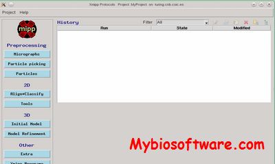
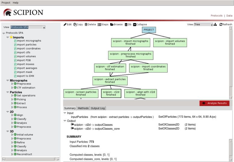
Scipion is an image processing framework to obtain 3D models of macromolecular complexes using Electron Microscopy (3DEM). It integrates several software packages and presents an unified interface for both biologists and developers.

::DEVELOPER

Biocomputing Unit – CNB

:: SCREENSHOTS

:: REQUIREMENTS

  • Linux
  • Xmipp

:: DOWNLOAD

  Scipion

:: MORE INFORMATION

Citation:

Scipion: a software framework toward integration, reproducibility and validation in 3D Electron Microscopy.
de la Rosa-Trevín JM, Quintana A, Del Cano L, Zaldívar A, Foche I, Gutiérrez J, Gómez-Blanco J, Burguet-Castell J, Cuenca-Alba J, Abrishami V, Vargas J, Otón J, Sharov G, Vilas JL, Navas J, Conesa P, Kazemi M, Marabini R, Sorzano CO, Carazo JM.
J Struct Biol. 2016 Apr 20. pii: S1047-8477(16)30079-X. doi: 10.1016/j.jsb.2016.04.010

A statistical approach to the initial volume problem in Single Particle Analysis by Electron Microscopy.
Sorzano CO, Vargas J, de la Rosa-Trevín JM, Otón J, álvarez-Cabrera AL, Abrishami V, Sesmero E, Marabini R, Carazo JM.
J Struct Biol. 2015 Mar;189(3):213-9. doi: 10.1016/j.jsb.2015.01.009.
Xmipp 3.1 – X-Windows-based Microscopy Image Processing Package

Xmipp 3.1

:: DESCRIPTION

Xmipp is a suite of image processing programs, primarily aimed at single-particle 3D electron microscopy.

::DEVELOPER

Biocomputing Unit

:: SCREENSHOTS

:: REQUIREMENTS

  •  Windows with Cygwin /Linux/MacOsX
  • Python

:: DOWNLOAD

 Xmipp

:: MORE INFORMATION

Citation

de la Rosa-Trevín JM, Otón J, Marabini R, Zaldívar A, Vargas J, Carazo JM, Sorzano CO.
Xmipp 3.0: an improved software suite for image processing in electron microscopy.
J Struct Biol. 2013 Nov;184(2):321-8. doi: 10.1016/j.jsb.2013.09.015. Epub 2013 Sep 26. PMID: 24075951.

C.O.S. Sorzano, R. Marabini, J.M. Carazo, J. Velazquez-Muriel, J.R. Bilbao-Castro, S.H.W. Scheres, J.M. Carazo and A. Pascual-Montano.
XMIPP: A new generation of an open-source image processing package for Electron Microscopy.
J Struct Biol. 2004 Nov;148(2):194-204.

ImageJ 2.2.0 – Image Processing & Analysis in Java

ImageJ 2.2.0

:: DESCRIPTION

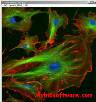
ImageJ is a public domain Java image processing program.ImageJ was designed with an open architecture that provides extensibility via Java plugins and recordable macros. Custom acquisition, analysis and processing plugins can be developed using ImageJ’s built-in editor and a Java compiler. User-written plugins make it possible to solve many image processing and analysis problems, from three-dimensional live-cell imaging, to radiological image processing, multiple imaging system data comparisons to automated hematology systems.

::DEVELOPER

ImageJ team

:: SCREENSHOTS

:: REQUIREMENTS

  • Windows / Linux / Mac OsX
  • Java

:: DOWNLOAD

ImageJ

:: MORE INFORMATION

Citation:

Abramoff, M.D., Magelhaes, P.J., Ram, S.J. “Image Processing with ImageJ“.
Biophotonics International, volume 11, issue 7, pp. 36-42, 2004.

ImgLib2 2-5.12.0 – Image Processing JAVA Library

ImgLib2 2-5.12.0

:: DESCRIPTION

ImgLib2 is a general-purpose, multidimensional image processing library.

::DEVELOPER

ImgLib2 team

:: SCREENSHOTS

N/A

:: REQUIREMENTS

  • Windows / Linux / MacOSX
  • Java

:: DOWNLOAD

  ImgLib2

:: MORE INFORMATION

Citation

Bioinformatics. 2012 Nov 15;28(22):3009-11. doi: 10.1093/bioinformatics/bts543. Epub 2012 Sep 8.
ImgLib2–generic image processing in Java.
Pietzsch T, Preibisch S, Tomancák P, Saalfeld S.

SPIDER 26.06 – Image processing system for Electron Microscopy

SPIDER 26.06

:: DESCRIPTION

SPIDER (System for Processing Image Data in Electron microscopy and Related fields) is an image processing system for electron microscopy, especially usefull for single-particle reconstruction.

::DEVELOPER

Wadsworth Bioinformatics Center

:: SCREENSHOTS

N/A

:: REQUIREMENTS

  • Linux

:: DOWNLOAD

SPIDER

:: MORE INFORMATION

Citation

Shaikh TR, Gao H, Baxter WT, Asturias FJ, Boisset N, Leith A, Frank J.
SPIDER image processing for single-particle reconstruction of biological macromolecules from electron micrographs.
Nat Protoc. 2008;3(12):1941-74. doi: 10.1038/nprot.2008.156. PMID: 19180078; PMCID: PMC2737740.

EBImage 4.35.0 – Image Processing and Analysis Toolkit for R

EBImage 4.35.0

:: DESCRIPTION

EBImage provides general purpose functionality for reading, writing, processing and analysis of images. Furthermore, in the context of microscopy-based cellular assays, EBImage offers tools to segment cells and extract quantitative cellular descriptors.

::DEVELOPER

Huber Group

:: SCREENSHOTS

N/A

:: REQUIREMENTS

  • Linux/ WIndows/MacOsX
  • R package
  • BioConductor

:: DOWNLOAD

 EBImage

:: MORE INFORMATION

Citation

Bioinformatics. 2010 Apr 1;26(7):979-81. doi: 10.1093/bioinformatics/btq046.
EBImage–an R package for image processing with applications to cellular phenotypes.
Pau G, Fuchs F, Sklyar O, Boutros M, Huber W.

AIM 0.82 – Automatic Image Processing for Microarrays

AIM 0.82

:: DESCRIPTION

AIM (Automatic Image Processing for Microarrays) is designed for uncalibrated microarray gridding and quantitative image analysis.Uncalibrated microarray image analysis supports integration of expression data from different sources and can improve reproducibility.

::DEVELOPER

Mathias Katzer

:: SCREENSHOTS

N/A

:: REQUIREMENTS

  • Linux

:: DOWNLOAD

AIM

:: MORE INFORMATION

Citation

M. Katzer and and F. Kummert and G. Sagerer:
Methods for Automatic Microarray Image Segmentation.
IEEE Transactions on Nano-Bioscience, 2003, pages 202-214

Fiji 20141125 – Image Processing Package based on ImageJ

Fiji 20141125

:: DESCRIPTION

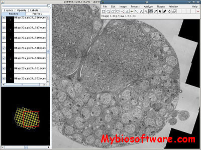
Fiji (Fiji is Just ImageJ) is an image processing package. It can be described as a distribution of ImageJ together with Java, Java 3D and a lot of plugins organized into a coherent menu structure.

::DEVELOPER

Fiji Team

:: SCREENSHOTS

:: REQUIREMENTS

  • Windows / Linux / MacOSX
  • Java

:: DOWNLOAD

Fiji

:: MORE INFORMATION

Publications introducing Fiji