Scipion 3.0 – An Image Processing Framework for 3D Electron Microscopy

Scipion 3.0

:: DESCRIPTION

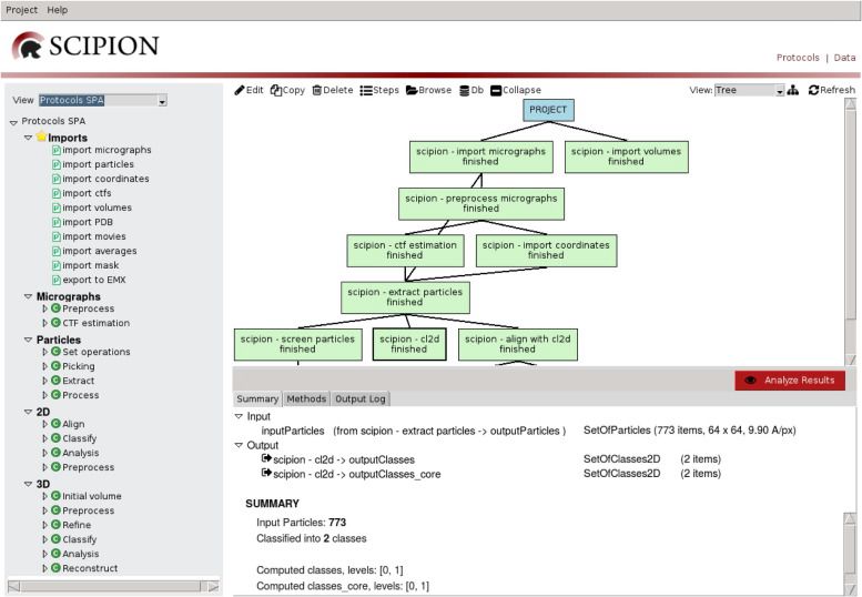
Scipion is an image processing framework to obtain 3D models of macromolecular complexes using Electron Microscopy (3DEM). It integrates several software packages and presents an unified interface for both biologists and developers.

::DEVELOPER

Biocomputing Unit – CNB

:: SCREENSHOTS

:: REQUIREMENTS

  • Linux
  • Xmipp

:: DOWNLOAD

  Scipion

:: MORE INFORMATION

Citation:

Scipion: a software framework toward integration, reproducibility and validation in 3D Electron Microscopy.
de la Rosa-Trevín JM, Quintana A, Del Cano L, Zaldívar A, Foche I, Gutiérrez J, Gómez-Blanco J, Burguet-Castell J, Cuenca-Alba J, Abrishami V, Vargas J, Otón J, Sharov G, Vilas JL, Navas J, Conesa P, Kazemi M, Marabini R, Sorzano CO, Carazo JM.
J Struct Biol. 2016 Apr 20. pii: S1047-8477(16)30079-X. doi: 10.1016/j.jsb.2016.04.010

A statistical approach to the initial volume problem in Single Particle Analysis by Electron Microscopy.
Sorzano CO, Vargas J, de la Rosa-Trevín JM, Otón J, álvarez-Cabrera AL, Abrishami V, Sesmero E, Marabini R, Carazo JM.
J Struct Biol. 2015 Mar;189(3):213-9. doi: 10.1016/j.jsb.2015.01.009.

Xmipp 3.1 – X-Windows-based Microscopy Image Processing Package

Xmipp 3.1

:: DESCRIPTION

Xmipp is a suite of image processing programs, primarily aimed at single-particle 3D electron microscopy.

::DEVELOPER

Biocomputing Unit

:: SCREENSHOTS

:: REQUIREMENTS

  •  Windows with Cygwin /Linux/MacOsX
  • Python

:: DOWNLOAD

 Xmipp

:: MORE INFORMATION

Citation

de la Rosa-Trevín JM, Otón J, Marabini R, Zaldívar A, Vargas J, Carazo JM, Sorzano CO.
Xmipp 3.0: an improved software suite for image processing in electron microscopy.
J Struct Biol. 2013 Nov;184(2):321-8. doi: 10.1016/j.jsb.2013.09.015. Epub 2013 Sep 26. PMID: 24075951.

C.O.S. Sorzano, R. Marabini, J.M. Carazo, J. Velazquez-Muriel, J.R. Bilbao-Castro, S.H.W. Scheres, J.M. Carazo and A. Pascual-Montano.
XMIPP: A new generation of an open-source image processing package for Electron Microscopy.
J Struct Biol. 2004 Nov;148(2):194-204.

sephaCe v3 – Segmentation of Adherent Biological Cell Boundaries

sephaCe v3

:: DESCRIPTION

sephaCe ( stands for “see phase of cells”,) is an open-source tool for automatic segmentation of adherent biological cell boundaries from brightfield microscopy images.

::DEVELOPER

Rehan Ali

:: SCREENSHOTS

sephaCe

:: REQUIREMENTS

  • Windows / MacOSX /Linux
  • Matlab

:: DOWNLOAD

 sephaCe

:: MORE INFORMATION

LAS X 3.7.4 – Lite version of the confocal software Leica Application Suite

LAS X 3.7.4

:: DESCRIPTION

Leica Application Suite X (LAS X) is the one software platform for all Leica microscopes: It integrates confocal, widefield, stereo, super-resolution, and light-sheet instruments from Leica Microsystems.

::DEVELOPER

Leica Microsystems

:: SCREENSHOTS

LASAFlite

:: REQUIREMENTS

  • Windows

:: DOWNLOAD

 LAS AF lite

:: MORE INFORMATION

Gelapp 1.2.7 – DNA & Prot Gel Analyzer

Gelapp 1.2.7

:: DESCRIPTION

Gelapp is an app that allows you to analyze gel (both agarose and PAGE) bands with a log graph automatically generated from the markers. It allows automatic detection and loading for future uses. Utilizing Gabor filter, this app allows users to improve detection of bands automatically. For poor quality images, users can click can manually draw a box on the desired band or click on the image to determine the specified location.

::DEVELOPER

Antibody & Product Development Team of BII-p53Lab ASTAR

:: SCREENSHOTS

:: REQUIREMENTS

  • iOS / Android

:: DOWNLOAD

Gelapp for iOS / for Android.

:: MORE INFORMATION

Citation

Nguyen PV, Ghezal A, Hsueh YC, Boudier T, Gan SK, Lee HK.
Optimal processing for gel electrophoresis images: Applying Monte Carlo Tree Search in GelApp.
Electrophoresis. 2016 Aug;37(15-16):2208-16. doi: 10.1002/elps.201600197. Epub 2016 Jul 4. PMID: 27251892.

cellxpress pro 1.4.2 – Cellular Phenotype Profiling Software Platform

cellxpress pro 1.4.2

:: DESCRIPTION

cellXpress is a cellular phenotype profiling software platform. The platform is designed for fast and high-throughput analysis of cellular phenotypes based on microscopy images. It is especially useful for large-scale profiling of cellular responses to pharmacological compounds, gene knockdowns, and/or toxic substances.

::DEVELOPER

the Loo Lab at the Bioinformatics Institute (BII), Singapore.

:: SCREENSHOTS

cellxpress

:: REQUIREMENTS

  • Windows/ Linux

:: DOWNLOAD

 cellxpress

 :: MORE INFORMATION

Citation

BMC Bioinformatics. 2013;14 Suppl 16:S4. doi: 10.1186/1471-2105-14-S16-S4. Epub 2013 Oct 22.
cellXpress: a fast and user-friendly software platform for profiling cellular phenotypes.
Laksameethanasan D, Tan R, Toh G, Loo LH.

bcfind 1.0 – Brain cell finder

bcfind 1.0

:: DESCRIPTION

bcfind is a tool for fully automated localization of soma in 3D mouse brain images acquired by confocal light sheet microscopy.

::DEVELOPER

bcfind team

:: SCREENSHOTS

N/A

:: REQUIREMENTS

  • Linux
  • Python
:: DOWNLOAD

:: MORE INFORMATION

Citation

Bioinformatics. 2014 Sep 1;30(17):i587-i593. doi: 10.1093/bioinformatics/btu469.
Large-scale automated identification of mouse brain cells in confocal light sheet microscopy images.
Frasconi P, Silvestri L, Soda P, Cortini R, Pavone FS, Iannello G.

ImageJ 2.2.0 – Image Processing & Analysis in Java

ImageJ 2.2.0

:: DESCRIPTION

ImageJ is a public domain Java image processing program.ImageJ was designed with an open architecture that provides extensibility via Java plugins and recordable macros. Custom acquisition, analysis and processing plugins can be developed using ImageJ’s built-in editor and a Java compiler. User-written plugins make it possible to solve many image processing and analysis problems, from three-dimensional live-cell imaging, to radiological image processing, multiple imaging system data comparisons to automated hematology systems.

::DEVELOPER

ImageJ team

:: SCREENSHOTS

:: REQUIREMENTS

  • Windows / Linux / Mac OsX
  • Java

:: DOWNLOAD

ImageJ

:: MORE INFORMATION

Citation:

Abramoff, M.D., Magelhaes, P.J., Ram, S.J. “Image Processing with ImageJ“.
Biophotonics International, volume 11, issue 7, pp. 36-42, 2004.

FISH Finder 0.27 – A High-Throughput Tool for Analyzing FISH Images

FISH Finder 0.27

:: DESCRIPTION

Fluorescence in situ hybridization (FISH) is used to study the organization and the positioning of specific DNA sequences within the cell nucleus.FISH Finder is a graphical software tool to automatically analyze FISH images that vary significantly.

::DEVELOPER

The Imaging Sciences Lab @ FSU

:: SCREENSHOTS

FISHFinder

:: REQUIREMENTS

  • Linux / Windows/ MacOsX
  • MatLab

:: DOWNLOAD

 FISH Finder

:: MORE INFORMATION

Citation:

Bioinformatics. 2011 Apr 1;27(7):933-8. doi: 10.1093/bioinformatics/btr053. Epub 2011 Feb 9.
FISH Finder: a high-throughput tool for analyzing FISH images.
Shirley JW1, Ty S, Takebayashi S, Liu X, Gilbert DM.