SyncX CT Aligner 1.0 – Image Alignment for Tomography Reconstruction from Synchrotron X-ray Microscopic Images

SyncX CT Aligner 1.0

:: DESCRIPTION

SyncX CT Aligner is a computer method to compensate for the vibration of the rotational holder by aligning neighboring X-ray images.

::DEVELOPER

Chang-Chieh Cheng, Ph.D.

:: SCREENSHOTS

SyncX

:: REQUIREMENTS

  • Windows/MacOsX

:: DOWNLOAD

 SyncX CT Aligner

:: MORE INFORMATION

Citation:

Image alignment for tomography reconstruction from synchrotron X-ray microscopic images.
Cheng CC, Chien CC, Chen HH, Hwu Y, Ching YT.
PLoS One. 2014 Jan 9;9(1):e84675. doi: 10.1371/journal.pone.0084675.

ScientiFig v2.99 – Build Publication-ready Scientific Figures

ScientiFig v2.99

:: DESCRIPTION

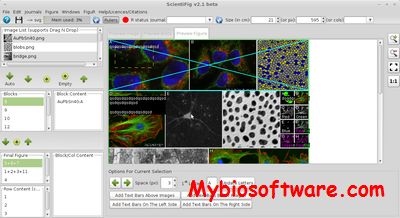
ScientiFig is a free tool to help you create, format or reformat scientific figures

::DEVELOPER

Benoit Aigouy (scientifig@gmail.com)

:: SCREENSHOTS

ScientiFig

:: REQUIREMENTS

  • Windows / Linux / MacOsX
  • Java
  • ImageJ / FIJI

:: DOWNLOAD

 ScientiFig

:: MORE INFORMATION

Citation

Nat Methods. 2013 Nov;10(11):1048. doi: 10.1038/nmeth.2692.
ScientiFig: a tool to build publication-ready scientific figures.
Aigouy B1, Mirouse V.

NucleusJ 1.0.3 – Quantifying 3D Images of Interphase Nuclei

NucleusJ 1.0.3

:: DESCRIPTION

NucleusJ is a simple and user-friendly ImageJ plugin dedicated to the characterization of nuclear morphology and chromatin organization in 3D.

::DEVELOPER

NucleusJ team

:: SCREENSHOTS

NucleusJ

:: REQUIREMENTS

  • Windows / Linux / MacOsX
  • Java
  • ImageJ

:: DOWNLOAD

 NucleusJ

 :: MORE INFORMATION

Citation

NucleusJ: an ImageJ plugin for quantifying 3D images of interphase nuclei.
Poulet A, Arganda-Carreras I, Legland D, Probst AV, Andrey P, Tatout C.
Bioinformatics. 2014 Nov 20. pii: btu774.

MorphoGraphX 1.1.1280 – Visualization and Processing of 3D Biological Datasets

MorphoGraphX 1.1.1280

:: DESCRIPTION

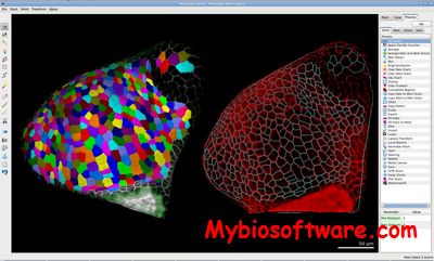
MorphoGraphX is a cross-platform, open-source application for the visualization and processing of 3D biological datasets.

::DEVELOPER

Richard S. Smith ,Systems Biology/Developmental Modeling

:: SCREENSHOTS

MorphoGraphX

::REQUIREMENTS

  • Linux

:: DOWNLOAD

 MorphoGraphX

:: MORE INFORMATION

Citation

Methods Mol Biol. 2014;1080:121-34. doi: 10.1007/978-1-62703-643-6_10.
Quantifying cell shape and gene expression in the shoot apical meristem using MorphoGraphX.
de Reuille PB1, Robinson S, Smith RS.

Daniel Kierzkowski et al.
Elastic Domains Regulate Growth and Organogenesis in the Plant Shoot Apical Meristem
Science 2 March 2012: Vol. 335 no. 6072 pp. 1096-1099

Fluorescence Spectraviewer 3.1.1 – Plot & Compare Spectra for iPhone/iPad

Fluorescence Spectraviewer 3.1.1

:: DESCRIPTION

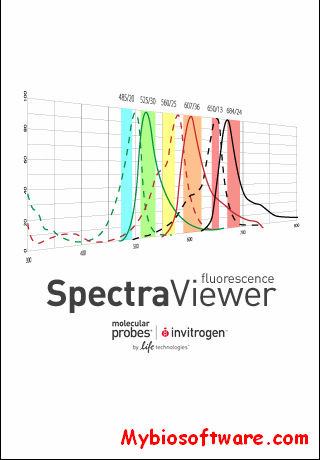
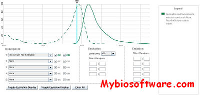
Fluorescence Spectraviewer is a software to plot and compare spectra, check the spectral compatibility for many fluorophores and email the configuration to yourself or others in the lab in a clear printable format.

::DEVELOPER

Thermo Fisher Scientific Inc.

:: SCREENSHOTS

:: REQUIREMENTS

  • iPhone /  iPad / Java

:: DOWNLOAD

Fluorescence Spectraviewer for iPhone

:: MORE INFORMATION

RegStatGel 1211 – Identifying differentially Expressed Proteins based on 2D Gel Images

RegStatGel 1211

:: DESCRIPTION

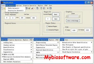
RegStatGel is a proteomic software for identifying differentially expressed proteins based on 2D gel images.

::DEVELOPER

Feng Li (feng.li@fda.hhs.gov)

:: SCREENSHOTS

RegStatGel

:: REQUIREMENTS

  • Windows
  • MatLab

:: DOWNLOAD

 RegStatGel

:: MORE INFORMATION

Citation

Bioinformation. 2011;6(10):389-90. Epub 2011 Aug 2.
RegStatGel: proteomic software for identifying differentially expressed proteins based on 2D gel images.
Li F, Seillier-Moiseiwitsch F.

Bio-Formats 6.6.1 – Java Library for Reading & Writing Biology Image File Formats

Bio-Formats 6.6.1

:: DESCRIPTION

Bio-Formats is a standalone Java library for reading and writing life sciences image file formats. It is capable of parsing both pixels and metadata for a large number of formats, as well as writing to several formats. See the table below for a complete list (click the headers to sort, and format names to see all information).

Bio-Formats’s primary purpose is to convert proprietary microscopy data into an open standard called the OME data model, particularly into the OME-TIFF file format.

::DEVELOPER

development teams at The Open Microscopy Environment,

:: SCREENSHOTS

:: REQUIREMENTS

  • Windows / Linux / Mac OsX
  • java

:: DOWNLOAD

Bio-Formats

:: MORE INFORMATION

NucType 1.5 – Measure Distances/Size of Chromosomes and analysis Karyotype

NucType 1.5

:: DESCRIPTION

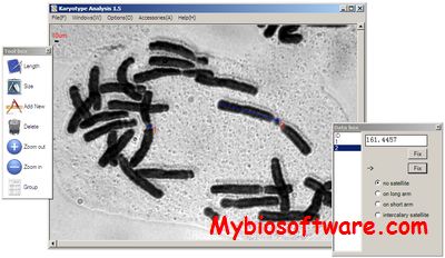
NucType is tool to measure distances/size of chromosomes, group the chromosomes and analysis the karyotype (karyotype asymmetry indices). Additionally it has a tool to measure the pollen or leaves.

::DEVELOPER

Yan Yu

:: SCREENSHOTS

NucType

:: REQUIREMENTS

  • Windows

:: DOWNLOAD

  NucType

:: MORE INFORMATION

Frida 1.1.0 – FRamework for Image Dataset Analysis

Frida 1.1.0

:: DESCRIPTION

Frida (FRamework for Image Dataset Analysis) is image analysis software. Frida was developed by the Johns Hopkins University Tissue Microarray Core Facility. Frida is open source and written in 100% Java. Frida makes use of functionality from the NIH’s ImageJ application.

::DEVELOPER

JHU Tissue MicroArray Core Facility

:: SCREENSHOTS

:: REQUIREMENTS

  • Linux / Mac OsX / Windows
  • Java

:: DOWNLOAD

Frida

:: MORE INFORMATION

Citation:

Cornish T, Morgan J, Gurel B, and De Marzo AM.
FrIDA: An open source framework for image dataset analysis.
Arch. Pathol. Lab. Med. 132:856 (2008). Originally presented at Advancing Practice, Instruction and Innovation Through Informatics: Pittsburgh, PA, 2007.