Scipion 3.0 – An Image Processing Framework for 3D Electron Microscopy

Scipion 3.0

:: DESCRIPTION

Scipion is an image processing framework to obtain 3D models of macromolecular complexes using Electron Microscopy (3DEM). It integrates several software packages and presents an unified interface for both biologists and developers.

::DEVELOPER

Biocomputing Unit – CNB

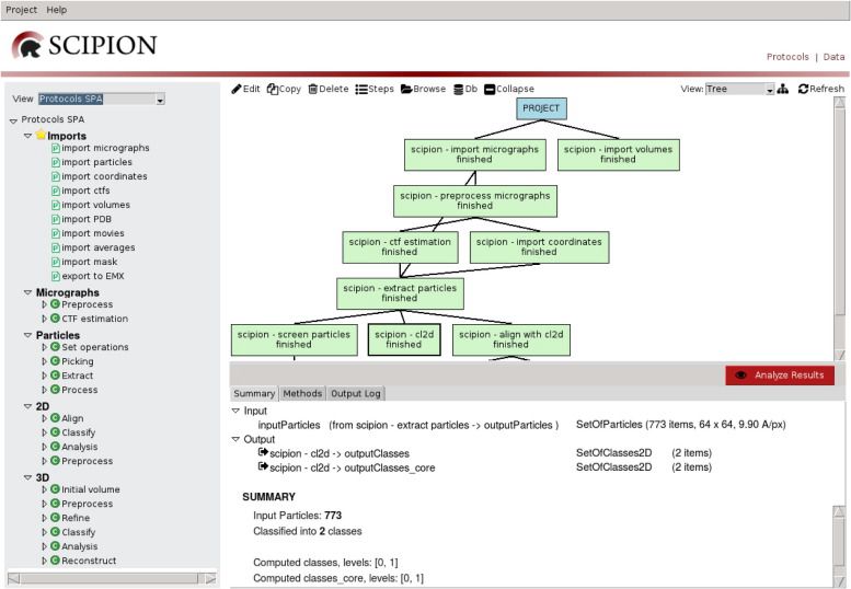
:: SCREENSHOTS

:: REQUIREMENTS

  • Linux
  • Xmipp

:: DOWNLOAD

  Scipion

:: MORE INFORMATION

Citation:

Scipion: a software framework toward integration, reproducibility and validation in 3D Electron Microscopy.
de la Rosa-Trevín JM, Quintana A, Del Cano L, Zaldívar A, Foche I, Gutiérrez J, Gómez-Blanco J, Burguet-Castell J, Cuenca-Alba J, Abrishami V, Vargas J, Otón J, Sharov G, Vilas JL, Navas J, Conesa P, Kazemi M, Marabini R, Sorzano CO, Carazo JM.
J Struct Biol. 2016 Apr 20. pii: S1047-8477(16)30079-X. doi: 10.1016/j.jsb.2016.04.010

A statistical approach to the initial volume problem in Single Particle Analysis by Electron Microscopy.
Sorzano CO, Vargas J, de la Rosa-Trevín JM, Otón J, álvarez-Cabrera AL, Abrishami V, Sesmero E, Marabini R, Carazo JM.
J Struct Biol. 2015 Mar;189(3):213-9. doi: 10.1016/j.jsb.2015.01.009.
SPIDER 26.06 – Image processing system for Electron Microscopy

SPIDER 26.06

:: DESCRIPTION

SPIDER (System for Processing Image Data in Electron microscopy and Related fields) is an image processing system for electron microscopy, especially usefull for single-particle reconstruction.

::DEVELOPER

Wadsworth Bioinformatics Center

:: SCREENSHOTS

N/A

:: REQUIREMENTS

  • Linux

:: DOWNLOAD

SPIDER

:: MORE INFORMATION

Citation

Shaikh TR, Gao H, Baxter WT, Asturias FJ, Boisset N, Leith A, Frank J.
SPIDER image processing for single-particle reconstruction of biological macromolecules from electron micrographs.
Nat Protoc. 2008;3(12):1941-74. doi: 10.1038/nprot.2008.156. PMID: 19180078; PMCID: PMC2737740.

EM-SURFER – Navigating 3D Electron Microscopy Maps

EM-SURFER

:: DESCRIPTION

EM-SURFER is a web platform for real-time electron microscopy database search. It compares isosurface shape of a query EM map against maps in the latest EMDB.

::DEVELOPER

Kihara Bioinformatics Laboratory

:: SCREENSHOTS

N/A

:: REQUIREMENTS

  • Web browser

:: DOWNLOAD

 NO

:: MORE INFORMATION

Citation

Navigating 3D electron microscopy maps with EM-SURFER.
Esquivel-Rodríguez J, Xiong Y, Han X, Guang S, Christoffer C, Kihara D.
BMC Bioinformatics. 2015 May 30;16:181. doi: 10.1186/s12859-015-0580-6.

Suprim 5.3.1 – Process Electron Microscopy Image

Suprim 5.3.1

:: DESCRIPTION

Suprim (SUpervisor for the PRocessing of IMages) is a flexible, modular software package intended for the processing of electron microscopy images is presented. The system consists of a set of image processing tools or filters, written in the C programming language, and a command line style user interface based on the UNIX shell. The pipe and filter structure of UNIX and the availability of command files in the form of shell scripts eases the construction of complex image processing procedures from the simpler tools. Implementation of a new image processing algorithm in SUPRIM may often be performed by construction of a new shell script, using already existing tools. Current, the package has been used for two- and three-dimensional image processing and reconstruction of macromolecules and other structures of biological interest.

::DEVELOPER

the Beckman Institute Imaging Technology Group

:: SCREENSHOTS

Command Lines

:: REQUIREMENTS

:: DOWNLOAD

Suprim

:: MORE INFORMATION

Citation

“SUPRIM: Easily Modified Image Processing Software”
John P. Schroeter and Jean-Pierre Bretaudiere
Journal of Structural Biology 116, 131-137 (1996)Article No. 0021. Volume 116, No. 1.