SPC Proteomics Tools 060107 – CDROM Image for SPC tools

SPC Proteomics Tools 060107

:: DESCRIPTION

SPC Proteomics Tools corom contains Seattle Proteome Center (SPC) – Proteomics Tools.

::DEVELOPER

Seattle Proteome Center

:: SCREENSHOTS

N/A

:: REQUIREMENTS

  • Windows / Mac OsX

:: DOWNLOAD

 SPC Proteomics Tools

:: MORE INFORMATION

this CD image contains an old version of the SPC tools. Please see the TPP wiki page for the most updated information.

GCellIQ 1.0 – Genomic Cell Image Quantitator

GCellIQ 1.0

:: DESCRIPTION

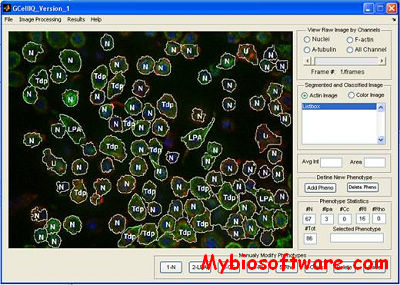
GCellIQ ( Genomic Cell Image Quantitator ) is user-friendly open-source software package that aims to provide an automated pipeline for processing large volumes of digital images generated from high-content screening (HCS), including those by RNAi or small compound screening. It’s suitable for batch processing large image dataset with little human interference.

::DEVELOPER

TMHRI Center for Bioengineering and Informatics

:: SCREENSHOTS

:: REQUIREMENTS

  • Windows / Linux / Mac OsX
  • MATLAB

:: DOWNLOAD

GCellIQ

:: MORE INFORMATION

Citation:

Li F, Zhou X, Zhu J, Ma J, Huang X, et al. (2007)
High content image analysis for human H4 neuroglioma cells exposed to CuO nanoparticles.
Bmc Biotechnology 7: p. 66.

VANO 1.741 – 3D Image Annotator

VANO 1.741

:: DESCRIPTION

VANO (Volume-object annotation system) is a System for 3D multicolor image stacks. VANO provides a well-coordinated way to annotate hundreds or thousands of 3D image objects. It combines 3D views of images and spread sheet neatly, and is just easy to manage 3D segmented image objects.

::DEVELOPER

Peng Lab @ JANELIA of HHMI

:: SCREENSHOTS

:: REQUIREMENTS

  • Windows / Linux / Mac OsX

:: DOWNLOAD

VANO

:: MORE INFORMATION

Citation:

Peng, H., Long, F., Myers, E.W.,
VANO: a volume-object image annotation system,”
Bioinformatics, Vol. 25, No. 5, pp. 695-697, 2009.

Gel Analyzer 2010a – 1D Gel Electrophoresis Image Analysis

Gel Analyzer 2010a

:: DESCRIPTION

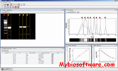
GelAnalyzer is a free 1D gel electrophoresis image analysis software .The process of gel image analysis is straightforward and covers all the main aspects of evaluation from automatic lane detection to precise molecular weight calculations. Many tools help you to improve the accuracy of gel evaluation, such as background subtraction methods and Rf calibration. These features make GelAnalyzer a real solution to gel image analysis in cost sensitive enviroments. With GelAnalyzer you don’t need expensive gel documentation system for simple image evaluation. You may obtain the gel image with a common digital camera, or even with your cell phone, and use GelAnalyzer to get the results in just a few seconds.

::DEVELOPER

lazarsoftware.com

:: SCREENSHOTS

:: REQUIREMENTS

  • Linux / Mac OsX / Windows
  • Java

:: DOWNLOAD

Gel Analyzer

:: MORE INFORMATION

By downloading, copying or installing any version of GelAnalyzer you accept to be bound by the End User Licence Agreement.

CMEIAS Color Segmentation 1.0 – Segment & Analyze Foreground Objects within Complex Images

CMEIAS Color Segmentation 1.0

:: DESCRIPTION

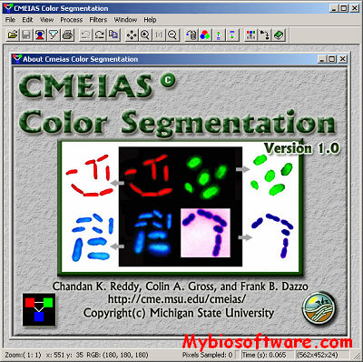
CMEIAS Color Segmentation is a free, improved computing technology designed to alleviate the laborious task of segmenting foreground objects of interest (e.g., microbes) from background in complex digital micrographs, as commonly encountered when preparing digital images of microbial populations and communities in environmental samples for quantitative image analysis.

::DEVELOPER

CMEIAS Team

:: SCREENSHOTS

:: REQUIREMENTS

  • Windows

:: DOWNLOAD

CMEIAS Color Segmentation

:: MORE INFORMATION

Citation

Colin A. Gross, Chandan K. Reddy and Frank B. Dazzo. 2009.
CMEIAS color segmentation: an improved computing technology to process color images for quantitative microbial ecology studies at single-cell resolution.
Microbial Ecology (2010) 59: 400-414. DOI:10.1007/s00248-009-9616-7.

LocusView 2.0 – Generate Images of Chromosomal Regions

LocusView 2.0

:: DESCRIPTION

LocusView is a program for generating images of chromosomal regions annotated with genomic features, experimental data, and analysis results.

:DEVELOPER

Tracey Petryshen, Andrew Kirby, and Mark Ainscow , The Broad Institute, Cambridge, MA

:: SCREENSHOTS

:: REQUIREMENTS

  • Linux / Mac OsX
  • Java

:: DOWNLOAD

LocusView

:: MORE INFORMATION

Citation

Please cite “T. Petryshen, A. Kirby, M. Ainscow, unpublished software” when using LocusView in publications

If you aren’t already registered, please send an email to locusview@broadinstitute.orgwith your name and email address, so that we can update you on new releases, bug fixes, etc.


Suprim 5.3.1 – Process Electron Microscopy Image

Suprim 5.3.1

:: DESCRIPTION

Suprim (SUpervisor for the PRocessing of IMages) is a flexible, modular software package intended for the processing of electron microscopy images is presented. The system consists of a set of image processing tools or filters, written in the C programming language, and a command line style user interface based on the UNIX shell. The pipe and filter structure of UNIX and the availability of command files in the form of shell scripts eases the construction of complex image processing procedures from the simpler tools. Implementation of a new image processing algorithm in SUPRIM may often be performed by construction of a new shell script, using already existing tools. Current, the package has been used for two- and three-dimensional image processing and reconstruction of macromolecules and other structures of biological interest.

::DEVELOPER

the Beckman Institute Imaging Technology Group

:: SCREENSHOTS

Command Lines

:: REQUIREMENTS

:: DOWNLOAD

Suprim

:: MORE INFORMATION

Citation

“SUPRIM: Easily Modified Image Processing Software”
John P. Schroeter and Jean-Pierre Bretaudiere
Journal of Structural Biology 116, 131-137 (1996)Article No. 0021. Volume 116, No. 1.

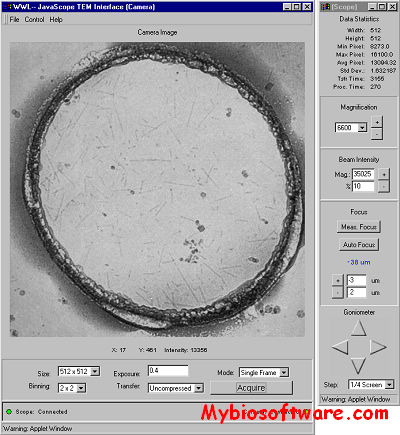
JavaScope 1.01 – Operate Philips CM200 Microscope & View Digital Images Remotely

JavaScope 1.01

:: DESCRIPTION

JavaScope is a web-based application designed to operate a Philips CM200 Transmission Electron Microscope (TEM) and to view digital images remotely. As a teaching and demonstration tool it can provide a “bulletproof” interface to the instrument for na?ve users and spectators. As a service tool it can be used by expert technicians to illustrate and explore specific problems with an instrument. This allows for a straightforward determination of the nature of a problem which benefits both customer and service provider. In this paper we will describe the implementation of the JavaScope application and discuss some of the benefits and disadvantages of the system.

::DEVELOPER

the Beckman Institute Imaging Technology Group

:: SCREENSHOTS

:: REQUIREMENTS

  • Linux / Mac OsX / Windows
  • Java

:: DOWNLOAD

JavaScope Source Code

:: MORE INFORMATION

If you have any questions, please mail javascope@itg.uiuc.edu.

TOPO2 – Create Transmembrane Proteins Images

TOPO2

:: DESCRIPTION

TOPO2 is an open source program written in Python for the creation of transmembrane protein 2D topology images. It makes no attempt to predict the TMDs that it displays. The user needs to supply that information. Residues of interest can be highlighted, if desired.

TOPO2 Web Version

::DEVELOPER

the Resource for Biocomputing, Visualization, and Informatics (RBVI) at UCSF

:: SCREENSHOTS

N/A

:: REQUIREMENTS

  • Linux /  MacOsX / Window
  • Python

:: DOWNLOAD

TOPO2

:: MORE INFORMATION

Contact Susan Jean Johns (email: johns@cgl.ucsf.edu) for more information about the software or if you are having problems with the site.