Visual3D 1.2 – Generate 3D Visualization of Fluorescence Microscopy Images

Visual3D 1.2

:: DESCRIPTION

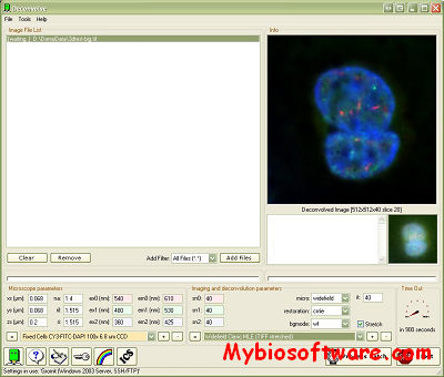
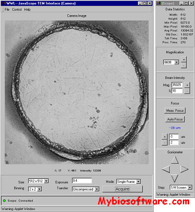
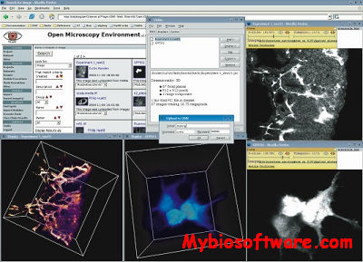
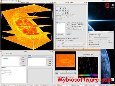
Visual3D uses OpenGL textures to generate a 3D visualization of confocal and wide field fluorescence microscopy images. Custom color palettes can be applied to grey value images. AVI movies can be created with a build in script engine.

::DEVELOPER

AMC, CMO, Amsterdam, The Netherlands – Contact: R.A.Hoebe -at- amc.nl

:: SCREENSHOTS

:: REQUIREMENTS

  • Windows

:: DOWNLOAD

Visual3D

:: MORE INFORMATION

This software is free to anyone, just for fun send author a postcard:
AMC, Cell biology & Histology CMO, Ron Hoebe, Room M3-106-1
Meibergdreef 15, 1105 AZ  Amsterdam, The Netherlands


Deconvolve 2.1 – Remote Batch Restorations of 3D/2D Fluorescence Images

Deconvolve 2.1

:: DESCRIPTION

Deconvolve is a Windows program for remote batch restorations of 3D/2D confocal and widefield fluorescence images using Huygens2 from SVI on a local or remote computer.

::DEVELOPER

AMC, CMO, Amsterdam, The Netherlands – Contact: R.A.Hoebe -at- amc.nl

:: SCREENSHOTS

:: REQUIREMENTS

  • Windows

:: DOWNLOAD

Deconvolve

:: MORE INFORMATION

This software is free to anyone, just for fun send author a postcard:
AMC, Cell biology & Histology CMO, Ron Hoebe, Room M3-106-1
Meibergdreef 15, 1105 AZ  Amsterdam, The Netherlands


top2maya – View 2D Atomic Force microscope Images as 3D Objects

top2maya

:: DESCRIPTION

top2maya is a script written in MEL (Maya Embedded Language) which runs in Alias|Wavefront’s Maya 2.5 and higher. It is a 3D visualization tool which analyzes images in 256 shades of gray and then displaces the individual pixels based on their color. It is a very effective tool for viewing 2D atomic force microscope images as 3D objects. As well as giving you a 3D image, it can also allow you to visualize multiple variables by doing the 3D displacement with one variable, for example the topography, then doing the shading on the new 3D object with another variable, for example surface friction.

::DEVELOPER

the Beckman Institute Imaging Technology Group

:: SCREENSHOTS

N/A

:: REQUIREMENTS

:: DOWNLOAD

geo.mel;  top2maya

:: MORE INFORMATION

N/A

ViewIt 4.23 – Multidimensional Image Processing Software

ViewIt 4.23

:: DESCRIPTION

ViewIt is basically multidimensional image processing software originally developed to handle data from magnetic resonance imaging. Some features of Viewit include:

Projection reconstruction algorithms for MRI data
Multidimensional Fourier transforms
Convolution operators for image smoothing and enhancement
Simple volume rendering functionality

::DEVELOPER

the Beckman Institute Imaging Technology Group

:: SCREENSHOTS

Command Lines

:: REQUIREMENTS

:: DOWNLOAD

ViewIt

:: MORE INFORMATION

Citation

“Viewit: A Software System for Multi-Dimensional Biomedical Image Processing, An alysis, and Visualization”
C.S. Potter and P.J. Moran,
SPIE Conference on Biomedical Image Processing III and T hree-Dimensional Microscopy, Vol 1660, February, 1992, pp. 767-773

tkImgDip 1.00b0 – Data Image Portal

tkImgDip 1.00b0

:: DESCRIPTION

tkImgDip is an image extension to Tcl/Tk for viewing arbitrary 1, 2, or 3 dimensional datasets and images as a pseudocolor (8-bit) image. The image represents portal over the data. The portal may be of any size, at any location over the data, and may apply magnification to the view of the data. The image displayed is rendered from the underlying data based on these and other parameters of the image portal.

::DEVELOPER

the Beckman Institute Imaging Technology Group

:: SCREENSHOTS

Command Lines

:: REQUIREMENTS

:: DOWNLOAD

tkImgDip

:: MORE INFORMATION

tkImgDip is no longer maintained by the ITG.


Suprim 5.3.1 – Process Electron Microscopy Image

Suprim 5.3.1

:: DESCRIPTION

Suprim (SUpervisor for the PRocessing of IMages) is a flexible, modular software package intended for the processing of electron microscopy images is presented. The system consists of a set of image processing tools or filters, written in the C programming language, and a command line style user interface based on the UNIX shell. The pipe and filter structure of UNIX and the availability of command files in the form of shell scripts eases the construction of complex image processing procedures from the simpler tools. Implementation of a new image processing algorithm in SUPRIM may often be performed by construction of a new shell script, using already existing tools. Current, the package has been used for two- and three-dimensional image processing and reconstruction of macromolecules and other structures of biological interest.

::DEVELOPER

the Beckman Institute Imaging Technology Group

:: SCREENSHOTS

Command Lines

:: REQUIREMENTS

:: DOWNLOAD

Suprim

:: MORE INFORMATION

Citation

“SUPRIM: Easily Modified Image Processing Software”
John P. Schroeter and Jean-Pierre Bretaudiere
Journal of Structural Biology 116, 131-137 (1996)Article No. 0021. Volume 116, No. 1.

JavaScope 1.01 – Operate Philips CM200 Microscope & View Digital Images Remotely

JavaScope 1.01

:: DESCRIPTION

JavaScope is a web-based application designed to operate a Philips CM200 Transmission Electron Microscope (TEM) and to view digital images remotely. As a teaching and demonstration tool it can provide a “bulletproof” interface to the instrument for na?ve users and spectators. As a service tool it can be used by expert technicians to illustrate and explore specific problems with an instrument. This allows for a straightforward determination of the nature of a problem which benefits both customer and service provider. In this paper we will describe the implementation of the JavaScope application and discuss some of the benefits and disadvantages of the system.

::DEVELOPER

the Beckman Institute Imaging Technology Group

:: SCREENSHOTS

:: REQUIREMENTS

  • Linux / Mac OsX / Windows
  • Java

:: DOWNLOAD

JavaScope Source Code

:: MORE INFORMATION

If you have any questions, please mail javascope@itg.uiuc.edu.

VisBio 3.40rc1 – Biological Visualization Tool

VisBio 3.40rc1

:: DESCRIPTION

VisBio is a biological visualization tool designed for easy visualization and analysis of multidimensional image data.VisBio provides 3D rendered views of multi-dimensional images that can be manipulated in 3D in real-time.

VisBio, using the remote OME Java API, can interact with OME directly for both reading and writing of image data.

::DEVELOPER

the Laboratory for Optical and Computational Instrumentation group

:: SCREENSHOTS

:: REQUIREMENTS

:: DOWNLOAD

VisBio ; Source code

:: MORE INFORMATION

VisBio is an open source project.

BQJavaLib 0.1 – Java Library for Matlab to Direct Access to the Bisque DataBase System.

BQJavaLib 0.1

:: DESCRIPTION

BQJavaLib is a Java library for Matlab that allows direct access to the Bisque DataBase system.

::DEVELOPER

The Center for Bio-Image Informatics

:: SCREENSHOTS

:: REQUIREMENTS

:: DOWNLOAD

BQJavaLib

:: MORE INFORMATION

Browse source on-line

BQMatlabToolbox 0.1 – Direct Access to the Bisque DataBase System

BQMatlabToolbox 0.1

:: DESCRIPTION

BQMatlabToolbox allows direct access to the Bisque DataBase system.

::DEVELOPER

The Center for Bio-Image Informatics

:: SCREENSHOTS

:: REQUIREMENTS

:: DOWNLOAD

BQMatlabToolbox

:: MORE INFORMATION

Browse source on-line

Exit mobile version