Raster3D 3.0-7 – Generate High Quality Raster Images of Proteins or other Molecules

Raster3D 3.0-7

:: DESCRIPTION

Raster3D is a set of tools for generating high quality raster images of proteins or other molecules. The core program renders spheres, triangles, cylinders, and quadric surfaces with specular highlighting, Phong shading, and shadowing. It uses an efficient software Z-buffer algorithm which is independent of any graphics hardware. Ancillary programs process atomic coordinates from PDB files into rendering descriptions for pictures composed of ribbons, space-filling atoms, bonds, ball+stick, etc. Raster3D can also be used to render pictures composed in other programs such as Molscript in glorious 3D with highlights, shadowing, etc. Output is to pixel image files with 24 bits of color information per pixel.

::DEVELOPER

Raster3D Team

:: SCREENSHOTS

N/A

:: REQUIREMENTS

  • MacOsx/Linux/Windows

:: DOWNLOAD

  Raster3D

:: MORE INFORMATION

Citation

Merritt, Ethan A. and Bacon, David J. (1997).
Raster3D: Photorealistic Molecular Graphics
Methods in Enzymology 277, 505-524.
DiagnoProt 1.4 – Discovery of new Molecules by Mass Spectrometry

DiagnoProt 1.4

:: DESCRIPTION

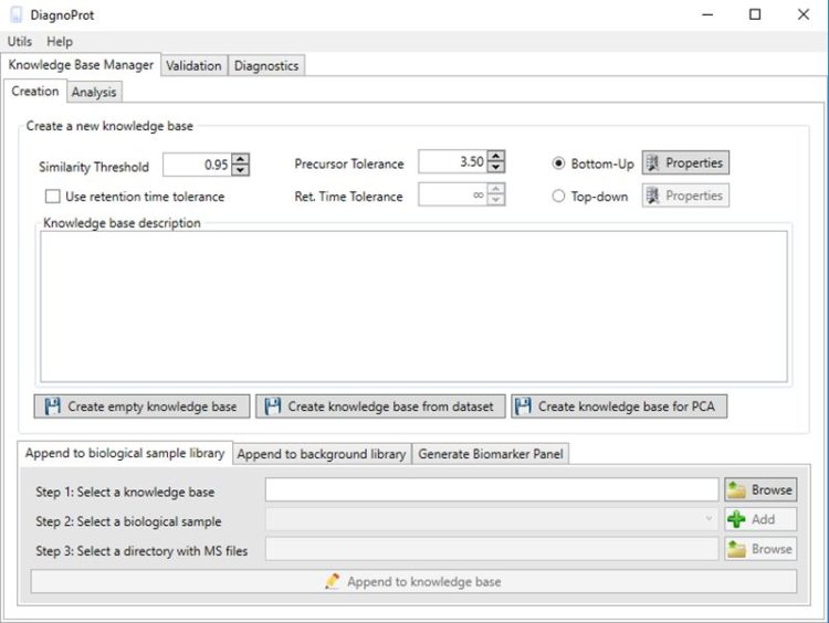
DiagnoProt is a tool that finds discriminative mass spectra among different biological conditions and ultimately performs spectral profiling classification of unknown conditions by comparing sets of tandem mass spectra (MS/MS).

::DEVELOPER

Laboratory for Computational and Strucutural Proteomics – Fiocruz

:: SCREENSHOTS

:: REQUIREMENTS

  • Windows
  • The .NET framework

:: DOWNLOAD

DiagnoProt 

:: MORE INFORMATION

Citation

Silva ARF, Lima DB, Leyva A, Duran R, Batthyany C, Aquino PF, Leal JC, Rodriguez JE, Domont GB, Santos MDM, Chamot-Rooke J, Barbosa VC, Carvalho PC.
DiagnoProt: a tool for discovery of new molecules by mass spectrometry.
Bioinformatics. 2017 Jun 15;33(12):1883-1885. doi: 10.1093/bioinformatics/btx093. PMID: 28186229.

PyWATER 1.0 – Find Conserved Water Molecules in Proteins by clustering

PyWATER 1.0

:: DESCRIPTION

PyWATER is a PyMOL plugin to find conserved water molecules in X-ray protein structure.

::DEVELOPER

PyWATER team

:: SCREENSHOTS

N/A

:: REQUIREMENTS

  • Windows/Linux/MacOs
  • Python
  • PyMOL

:: DOWNLOAD

 PyWATER

:: MORE INFORMATION

Citation

PyWATER: A PyMOL plugin to find conserved water molecules in proteins by clustering.
Patel H, Grüning BA, Günther S, Merfort I.
Bioinformatics. 2014 Jul 1. pii: btu424

iMolview 1.9.5 – iPhone, iPad and Android App for Browsing Protein, DNA & Drug Molecules in 3D

iMolview 1.9.5

:: DESCRIPTION

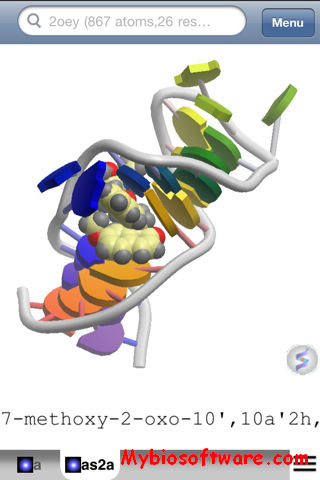
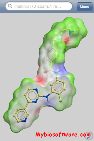
iMolview is an app for the iPhone , iPad and Android that lets you browse protein, DNA, and drug molecules in 3D. The app has a direct link to the Protein Data Bank (PDB) and DrugBank and has a fast and easy to use interface. Touching the molecules via the screen allows you to interact immediately with the 3D structures in a unique way. You can zoom in and out, rotate, spin, pan, and clip the 3D molecules with your finger tips in ways that are impossible using a traditional mouse and desktop computer.

::DEVELOPER

Molsoft LLC.

:: SCREENSHOTS

:: REQUIREMENTS

  • IPhone / IPad / Android

:: DOWNLOAD

iMolview

:: MORE INFORMATION

Avogadro2 1.90 – Molecule Editor & Visualizer

Avogadro2 1.90

:: DESCRIPTION

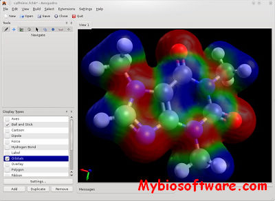
Avogadro is an advanced molecule editor and visualizer designed for cross-platform use in computational chemistry, molecular modeling, bioinformatics, materials science, and related areas. It offers flexible high quality rendering and a powerful plugin architecture.

::DEVELOPER

Avogadro team

:: SCREENSHOTS

:: REQUIREMENTS

  • Windows / MacOsX /  Linux

:: DOWNLOAD

Avogadro

:: MORE INFORMATION

Citation

J Cheminform. 2012 Aug 13;4(1):17. doi: 10.1186/1758-2946-4-17.
Avogadro: an advanced semantic chemical editor, visualization, and analysis platform.
Hanwell MD, Curtis DE, Lonie DC, Vandermeersch T, Zurek E, Hutchison GR.

Layers – An Algorithm that Peels Molecule as Layers

Layers

:: DESCRIPTION

Layers is a molecular surface peeling algorithm and its applications to analyze protein structures.

::DEVELOPER

Computational Structural Biology Lab , Indian Institute of Technology Kharagpur

:: SCREENSHOTS

N/A

:: REQUIREMENTS

  • Web browser

:: DOWNLOAD

  NO

:: MORE INFORMATION

Citation

Layers: A molecular surface peeling algorithm and its applications to analyze protein structures.
Karampudi NB, Bahadur RP.
Sci Rep. 2015 Nov 10;5:16141. doi: 10.1038/srep16141.

ICM-Browser 3.9-2b – Molecules & Sequence Alignments Visualization

ICM-Browser 3.9-2b

:: DESCRIPTION

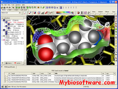
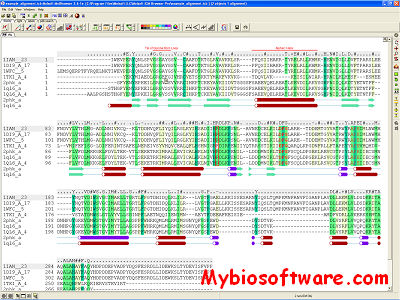
ICM-Browser provides a biologist or a chemist with direct access to the treasures of structural biology and protein families.It reads a variety of file formats directly from the database web-sites including: PDB, chemical, electron density maps, sequence and alignment files. ICM-Browser provides a rich professional molecular graphics environment with powerful representations of proteins, DNA and RNA, and multiple sequence alignments.

When you purchase the license ICM-Browser will become ICM-Browser-Pro.ICM-Browser-Pro is a high quality visualizer and annotator for three dimensional molecular structures, sequences, alignments, chemical spreadsheets and biological data. It allows you to read data from multiple file formats, annotate the data, and write multi-slide documents in a single small cross-platform file. ICM Browser Pro is well suited for creating, storing and sharing structural, biological and chemical information. The files can then be opened and viewed with the free ICM Browser.

::DEVELOPER

Molsoft LLC.

:: SCREENSHOTS

:: REQUIREMENTS

  • Windows/Linux/Mac OS X

:: DOWNLOAD

ICM-Browser ; Manual

:: MORE INFORMATION

VMD 1.9.4 – Molecular Graphics Viewer

VMD 1.9.4

:: DESCRIPTION

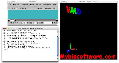
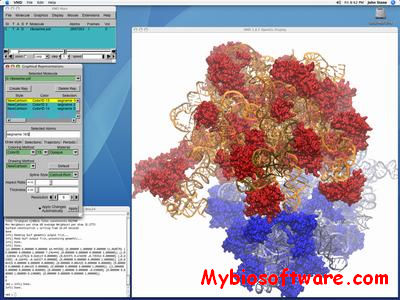
VMD is designed for modeling, visualization, and analysis of biological systems such as proteins, nucleic acids, lipid bilayer assemblies, etc. It may be used to view more general molecules, as VMD can read standard Protein Data Bank (PDB) files and display the contained structure. VMD provides a wide variety of methods for rendering and coloring a molecule: simple points and lines, CPK spheres and cylinders, licorice bonds, backbone tubes and ribbons, cartoon drawings, and others. VMD can be used to animate and analyze the trajectory of a molecular dynamics (MD) simulation. In particular, VMD can act as a graphical front end for an external MD program by displaying and animating a molecule undergoing simulation on a remote computer.

::DEVELOPER

Theoretical and Computational Biophysics group at the University of Illinois.

:: SCREENSHOTS

:: REQUIREMENTS

:: DOWNLOAD

VMD

:: MORE INFORMATION

Humphrey, W., Dalke, A. and Schulten, K., “VMD – Visual Molecular Dynamics”, J. Molec. Graphics, 1996, vol. 14, pp. 33-38.

DOCK 6.9 – Docking Molecules to each other

DOCK 6.9

:: DESCRIPTION

DOCK addresses the problem of “docking” molecules to each other. In general, “docking” is the identification of the low-energy binding modes of a small molecule, or ligand, within the active site of a macromolecule, or receptor, whose structure is known. A compound that interacts strongly with, or binds, a receptor associated with a disease may inhibit its function and thus act as a drug. Solving the docking problem computationally requires an accurate representation of the molecular energetics as well as an efficient algorithm to search the potential binding modes.

::DEVELOPER

DOCK Team

:: SCREENSHOTS

N/A

:: REQUIREMENTS

:: DOWNLOAD

DOCK

:: MORE INFORMATION

Lang, P.T., Brozell, S.R., Mukherjee, S., Pettersen, E.T., Meng, E.C., Thomas, V., Rizzo, R.C., Case, D.A., James, T.L., Kuntz, I.D.
DOCK 6: Combining Techniques to Model RNA-Small Molecule Complexes.
RNA 15:1219-1230, 2009.