Dapple 0.88pre4
:: DESCRIPTION
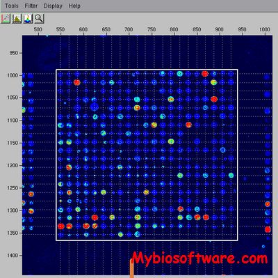
Dapple is a program for quantitating spots on a two-color DNA microarray image. Given a pair of images from a comparative hybridization, Dapple finds the individual spots on the image, evaluates their qualities, and quantifies their total fluorescent intensities.
Dapple is designed to work with microarrays on glass. The spot-finding techniques used are robust to uneven spot sizes and positional deviations caused by “wobbling” of the arraying robot, as well as image noise and artifacts. As long as your spots are consistently circular, Dapple has a good chance of finding them accurately.
::DEVELOPER
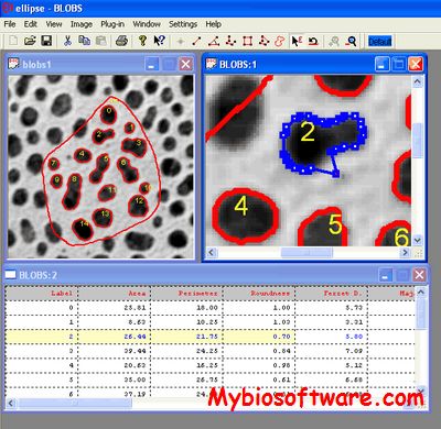
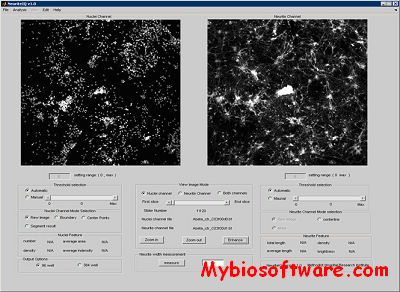
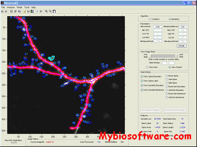
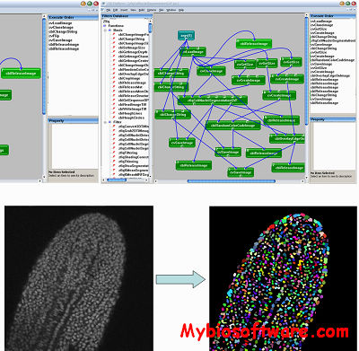
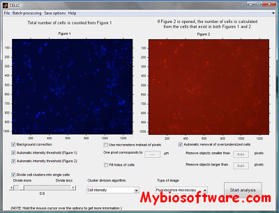
:: SCREENSHOTS

:: REQUIREMENTS
- Linux / Mac OsX
- FFTW library
- GCC
- Qt toolkit
:: DOWNLOAD

:: MORE INFORMATION
Citation:
J. Buhler, T. Ideker, D. Haynor, “Dapple: Improved Techniques for Finding Spots on DNA Microarrays”, University of Washington Department of Computer Science & Engineering Technical Report UW-CSE-2000-08-05, (2000) Supplement.